Clinical Cases
Clinical Case Analysis: Oral Ossifying Fibroma
Clinical Diagnosis, Surgical Treatment, and Histopathological Confirmation
Progressively growing oral lesions are a common diagnostic challenge in clinical practice. Although some lesions may initially appear benign, their clinical presentation requires a thorough evaluation to rule out more significant pathologies.
In this clinical case, we present the diagnosis and treatment of an oral ossifying fibroma, highlighting the importance of early diagnosis, appropriate surgical management, and histopathological confirmation.

Implante dental con regeneración ósea guiada
La implantología moderna permite reemplazar piezas dentales perdidas de forma segura, estética y duradera.
En este caso clínico, el Dr. Marco C. Moreno —cirujano oral y especialista en implantología— comparte el proceso completo de colocación de un implante con regeneración ósea guiada, desde el diagnóstico hasta el resultado final.
Clinical Cases
Clinical Case Analysis: Oral Ossifying Fibroma
Clinical Diagnosis, Surgical Treatment, and Histopathological Confirmation.
Progressively growing oral lesions are a common diagnostic challenge in clinical practice. Although some lesions may initially appear benign, their clinical presentation requires a thorough evaluation to rule out more significant pathologies.
In this clinical case, we present the diagnosis and treatment of an oral ossifying fibroma, highlighting the importance of early diagnosis, appropriate surgical management, and histopathological confirmation.
Initial Clinical Presentation
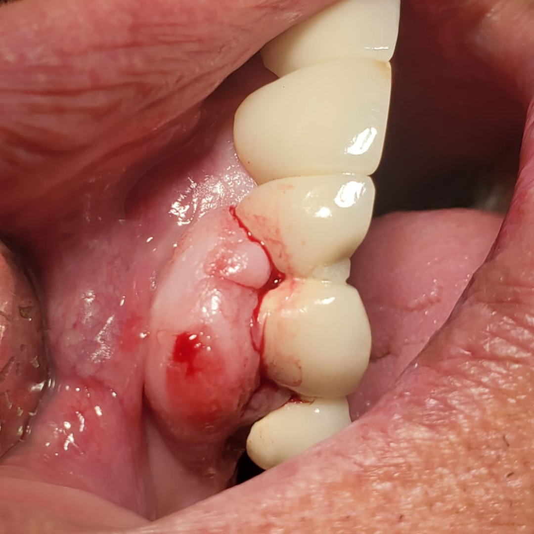
An exophytic lesion was identified on the oral mucosa, demonstrating progressive growth, an irregular surface, ulcerated areas, and noticeable epithelial changes.
During the clinical examination, bleeding upon contact and irregular lesion borders were observed, findings that warranted further diagnostic evaluation.
Initial Clinical Presentation
An exophytic lesion was identified on the oral mucosa, demonstrating progressive growth, an irregular surface, ulcerated areas, and noticeable epithelial changes.
During the clinical examination, bleeding upon contact and irregular lesion borders were observed, findings that warranted further diagnostic evaluation.
Surgical Procedure
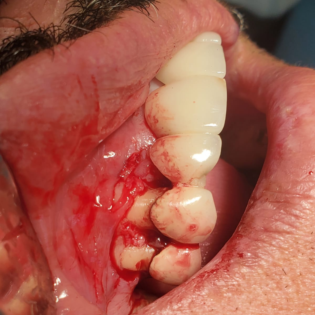
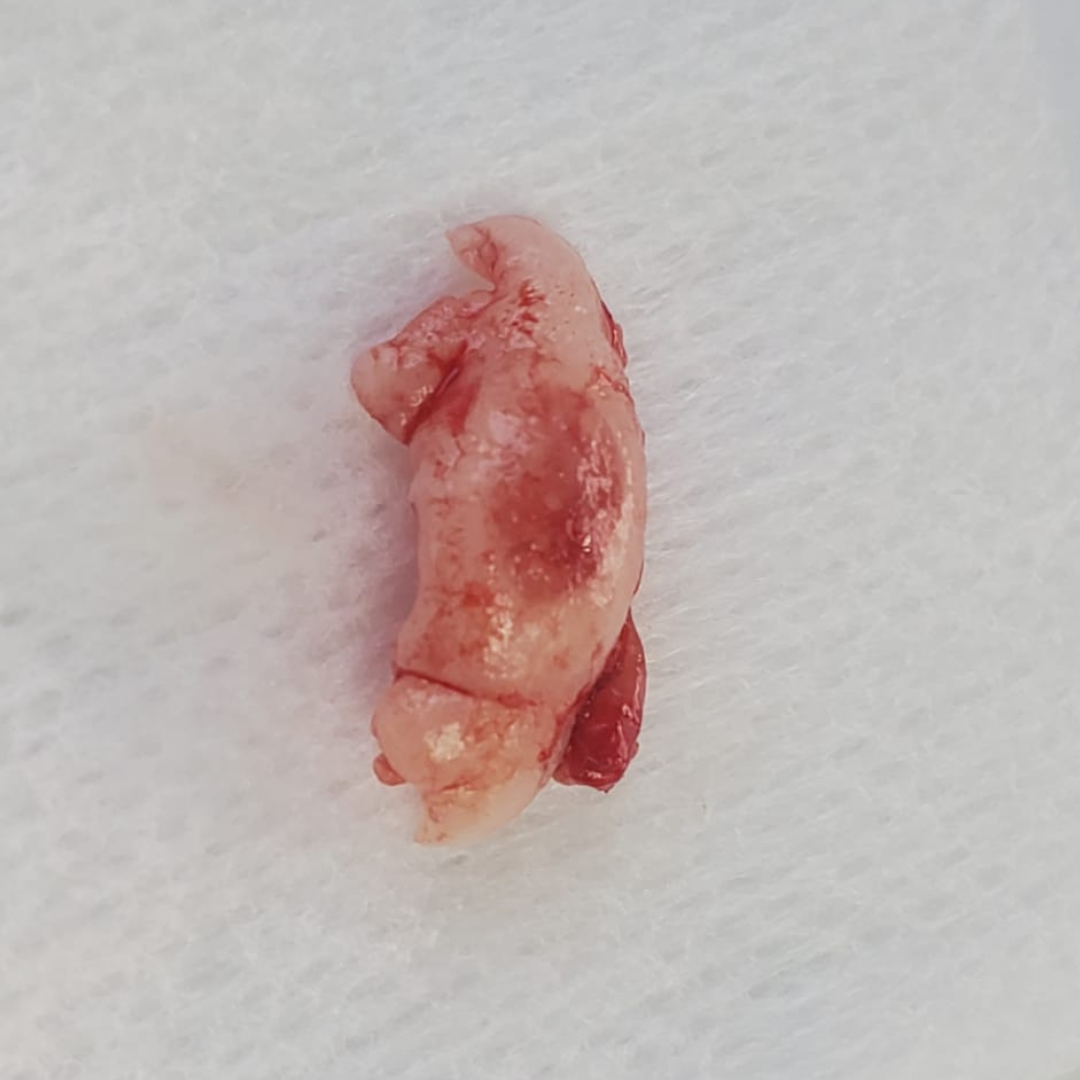
Following the clinical diagnosis, the lesion was completely removed through surgical excision while preserving healthy surrounding tissues and maintaining appropriate surgical margins.
The objective of the procedure was to eliminate the pathological tissue and reduce the risk of recurrence.
Early treatment plays a critical role in achieving the best long-term outcome.
Surgical Procedure
Following the clinical diagnosis, the lesion was completely removed through surgical excision while preserving healthy surrounding tissues and maintaining appropriate surgical margins.
The objective of the procedure was to eliminate the pathological tissue and reduce the risk of recurrence.
Early treatment plays a critical role in achieving the best long-term outcome.
Histopathological Analysis
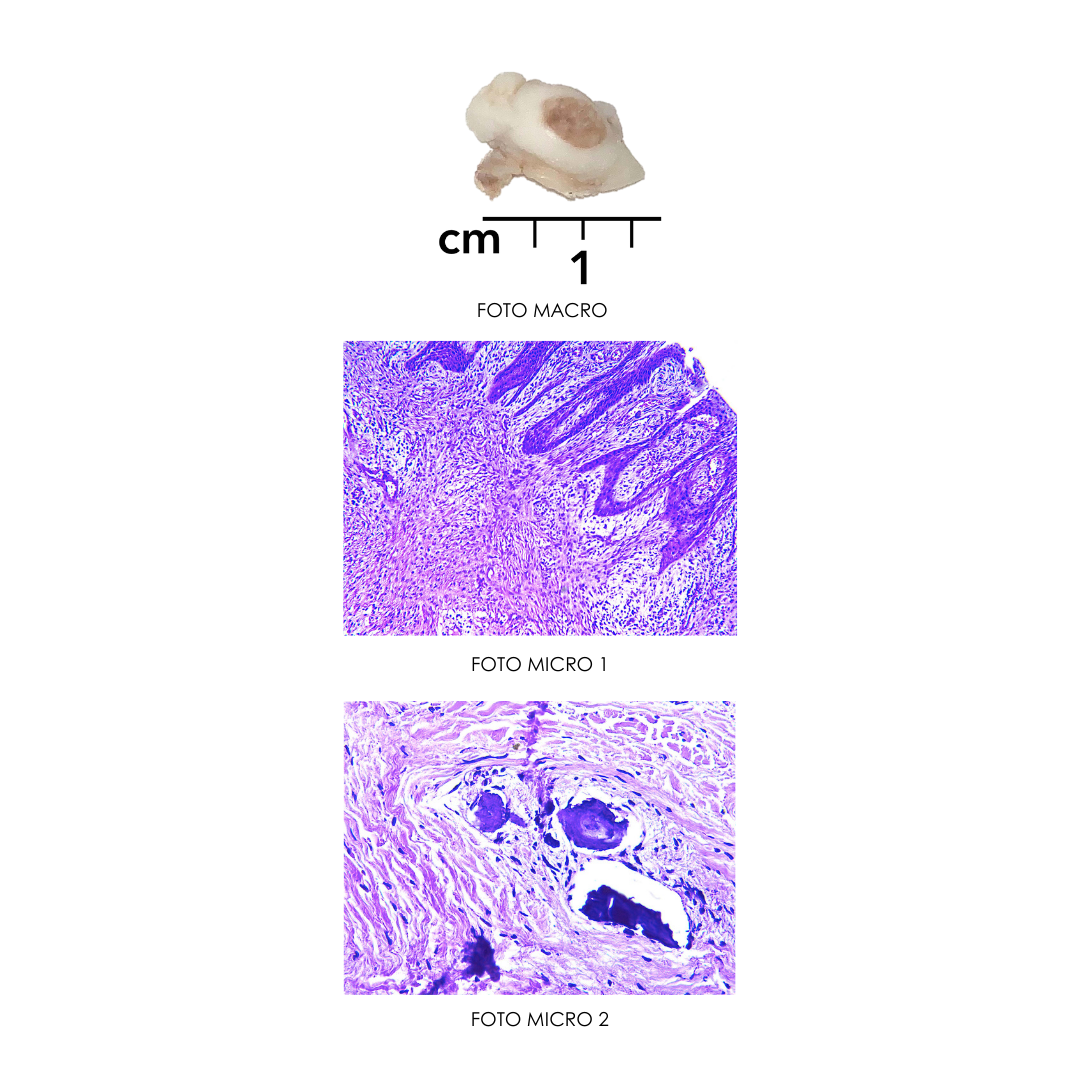
A biopsy and microscopic evaluation were performed to establish a definitive diagnosis.
Histopathological examination provided detailed information about the tissue architecture and cellular characteristics, eliminating diagnostic uncertainty.
📌 Final Diagnosis: Oral Ossifying Fibroma
Surgical Closure & Recovery
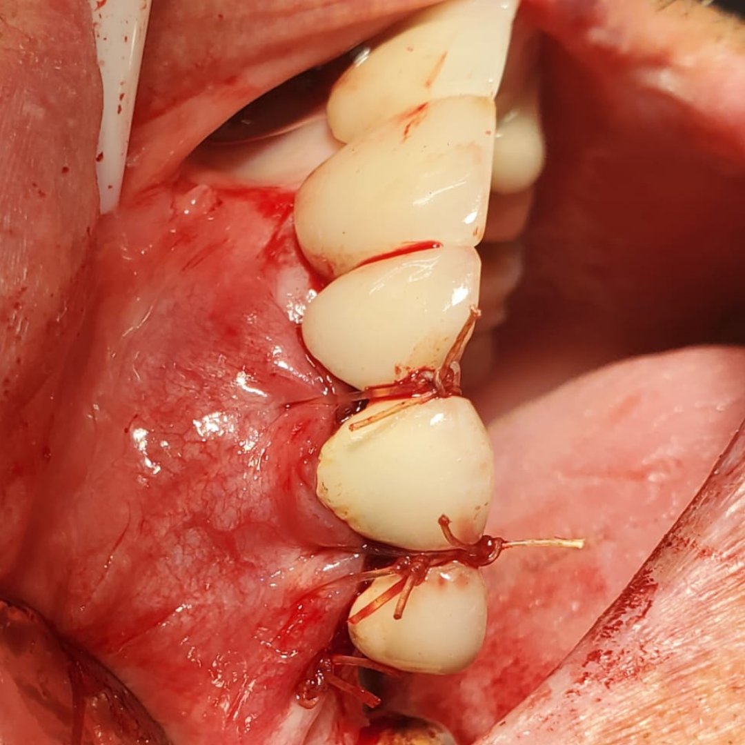
Following lesion removal, the surgical site was closed with sutures to ensure proper tissue adaptation and promote optimal healing.
Postoperative follow-up is essential to monitor healing and ensure a successful recovery.
Long-term follow-up is an important part of treatment.
Histopathological Analysis
A biopsy and microscopic evaluation were performed to establish a definitive diagnosis.
Histopathological examination provided detailed information about the tissue architecture and cellular characteristics, eliminating diagnostic uncertainty.
📌Final Diagnosis: Oral Ossifying Fibroma
Surgical Closure & Recovery
Following lesion removal, the surgical site was closed with sutures to ensure proper tissue adaptation and promote optimal healing.
Postoperative follow-up is essential to monitor healing and ensure a successful recovery.
Long-term follow-up is an important part of treatment.
Why Professional Evaluation Matters
Although many oral lesions are benign, professional evaluation is essential to obtain an accurate diagnosis and determine the appropriate treatment.
Early detection helps prevent complications and provides patients with greater peace of mind.
Some lesions may remain stable or resolve naturally, while others require surgical intervention. An evaluation by an experienced oral surgeon ensures safe treatment and appropriate follow-up care.
📞 Schedule Your Evaluation
Have you noticed a lump, lesion, or unusual change in your mouth or gums?
Early diagnosis can make all the difference.
Schedule your consultation with Dr. Marco Moreno, Certified Oral Surgeon in Tijuana.
📲 WhatsApp or Call: (619) 432-0502 | (664) 977-5558
Why Professional Evaluation Matters
Although many oral lesions are benign, professional evaluation is essential to obtain an accurate diagnosis and determine the appropriate treatment.
Early detection helps prevent complications and provides patients with greater peace of mind.
Some lesions may remain stable or resolve naturally, while others require surgical intervention. An evaluation by an experienced oral surgeon ensures safe treatment and appropriate follow-up care.
📞 Schedule Your Evaluation
Have you noticed a lump, lesion, or unusual change in your mouth or gums?
Early diagnosis can make all the difference.
Schedule your consultation with Dr. Marco Moreno, Certified Oral Surgeon in Tijuana.
📲 WhatsApp or Call: (619) 432-0502 | (664) 977-5558